Chapter outline

  1. Overview of cell biology
  2. Inside the cell:
    • Cell membrane, cytoplasm
    • Organelles
  3. Outside the cell:
  4. Neither in nor out:
    • Cell division
    • Cell death
    • Cell junctions

cell

Philosophy of cell biology

The cell is the basic unit of life. It is the smallest thing that we call living (without arguing about viruses), and the human body is made of 10 trillion of them. We started off as a single cell, and its the purpose of this class to learn a little bit about how that one cell developed into the trillion-celled organism you are today. Nearly all of the instructions for making trillions of cells-- how to make them, when to make them, where to make them-- are found within that single cell.


Different cells have different functions, even though almost every cell in one person has the exact same DNA. To become different from one another, cells will express different DNA. This is an impotant process called differentiation, which is really no more complicated in concept than cells going from looking like boring generic cells to looking and behaving differently from other cells. We give different names to cells as they make these changes. For instance, a stem cell that gives rise to, oh lets say, a smurf, would be named a Smurfal Stem Cell. As the Smurfal Stem Cells divide and differentiate into cells that make a smurf, the cells actually making the smurf would be named Smurfoblasts, and when the Smurf was finished, the cells inside that Smurf would be named Smurfocytes. The stem cells found in a tissue called mesenchyme are named Mesenchymal Stem Cells, the cells that make dentin are named odontoblasts, the cells within mature cementum are named cementocytes


The aim of this chapter is to review aspects of a cell biology class that come up in this textbook, and not much more. I assume you have covered cell biology in a pre-requisite to this class. We need to review the parts of a cell that help us to explain difficult concepts like differentiation and development. If you find you need more than a quick refresher, here are some link from the NIH, who have a number of very useful publications and videos (for free), such as: Two other useful (and free) eBooks you may wish to download are from OpenStax:


membrane

Cell membrane and cytoplasm

Every human cell is surrounded by the plasma membrane. The plasma membrane separates the cell from its environment, and allows certain materials to enter and leave the cell. Phospholipids and cholesterol form a barrier that separates the cell from the external environment, and allows us to point at that cell and call it a thing. Trans-membrane proteins span the plasma membrane, and decide what goes in or out. These proteins can also receive signals from other cells and relay that information to the inside of the cell, perhaps even to the nucleus.


The cytoplasm is the gelatinous filling of a cell, and is sometimes referred to an Intra-Cellular Fluid (ICF). Cytoplasm includes nutrients and electrolytes absorbed from the Extra-Cellular Fluid (ECF), or the fuid surrounding all cells. In addition, the cytoplasm includes a number of proteins and glycoproteins synthesized by the cell. These molecules may have other important functions, but they attract water from the ECF. The end result is that cells have a gelatinous filling, rather than a watery one. This gelatinous filling is filled with a number of organelles, similar to the way my grandmother's jello salad contained grapes, raisins and other food or food-like substances that did not in any way turn that jello into an actual salad.

cell

The nucleus

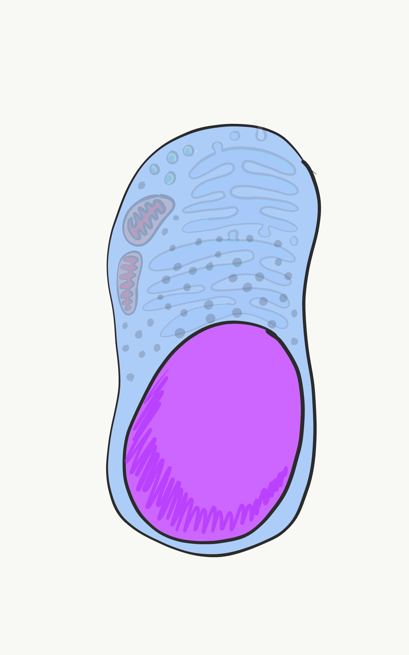
The nucleus contains a cell's DNA. This DNA is the instructions for making all of the proteins inside of and outside of a cell. It is also the instructions for when and where to make these proteins. For instance, epithelial cells of the oral mucosa will not make collagen or enzymes that secrete calcium and phosphate into the ECM. On the other hand, epithelial cells that differentiate into ameloblasts will make these proteins, by expressing their genes, after being told to do so by other cells called neural crest cells.


All cells in the human body have the same DNA (with a few exceptions). However, different cells will express different DNA at different times. DNA can be divided into 2 basic types. There are genes, each gene is more-or-less the instructions for a single protein. The rest of DNA folds up into unique shapes that provides instructions for when and where to express these genes. The latter is referred to as non-coding DNA. For DNA expression to occur, proteins called transcription factors (with help from special small molecules) bind to regulatory regions of DNA and inititate the transcription of that gene into mRNA, which leaves the nucleus to be translated into protein. Other transcription factors may temporarily inhibit the expression of genes.


histone
Transcription factors turn on and turn off genes quickly, in reponse to changes in a cells environment. But when cells differentiate, they shut down genes they don't need more permanently. Rather that relying on the on and off switches of transcription factors binding to regulatory DNA regions, these inactivated genes will be methylated, packed up around histones, and stored.


We have 46 molecules of DNA in the nucleus-- they are freakishly long molecules, but only 46 in number-- 23 maternal, and 23 paternal. During mitosis, these 46 molecules are packaged up tightly into 46 chromosomes (times 2) that can be seen under the light microscope. The rest of the time, DNA is mostly unwound (un-needed instructions are wound around histones, the rest are free to be transcribed), and fills up the nucleus in a way that doesn't look very exciting. We call that DNA chromatin, and functionally it is by far the more exciting of the two forms.

cell

Ribosomes

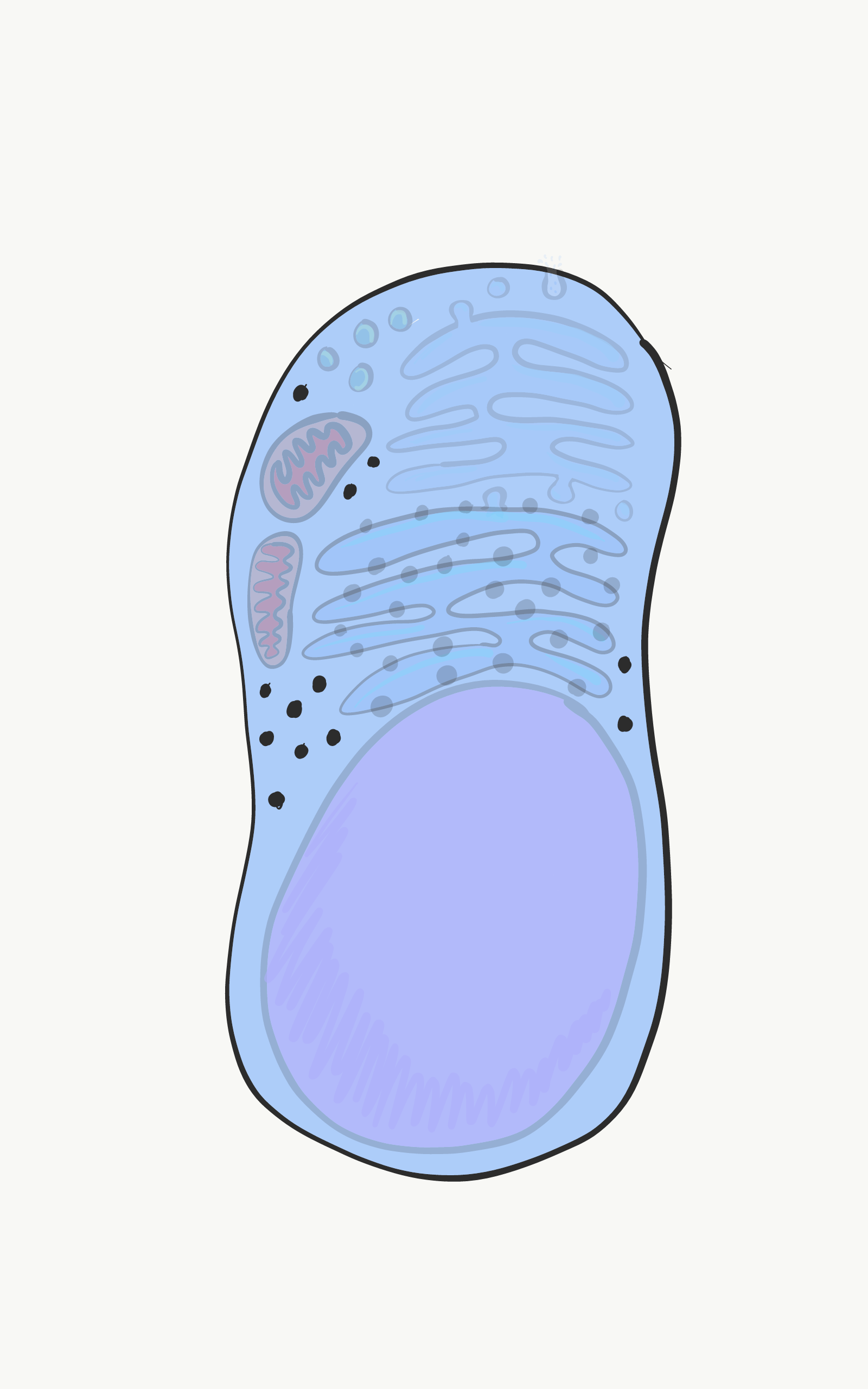
Visible throughout the cytoplasm are small specks made of protein and RNA called ribosomes. These structures translate mRNA instructions that came from the nucleus into protein. Groups of three mRNA nucleotides, called codons, instruct the ribosome which amino acid to add to a protein. Like the regulatory regions of DNA, the RNA found in the ribosome itself-- not the mRNA instructions but ribosomal rRNA, is a type of RNA that folds up into specific shapes that, along with the ribosomal proteins, perform the enzymatic reaction of translation. Free-floating ribosomes in the cytoplasm synthesize proteins that remain in the cytoplasm, such as keratin or enzymes that mediate apoptosis.

cell

Mitochondria

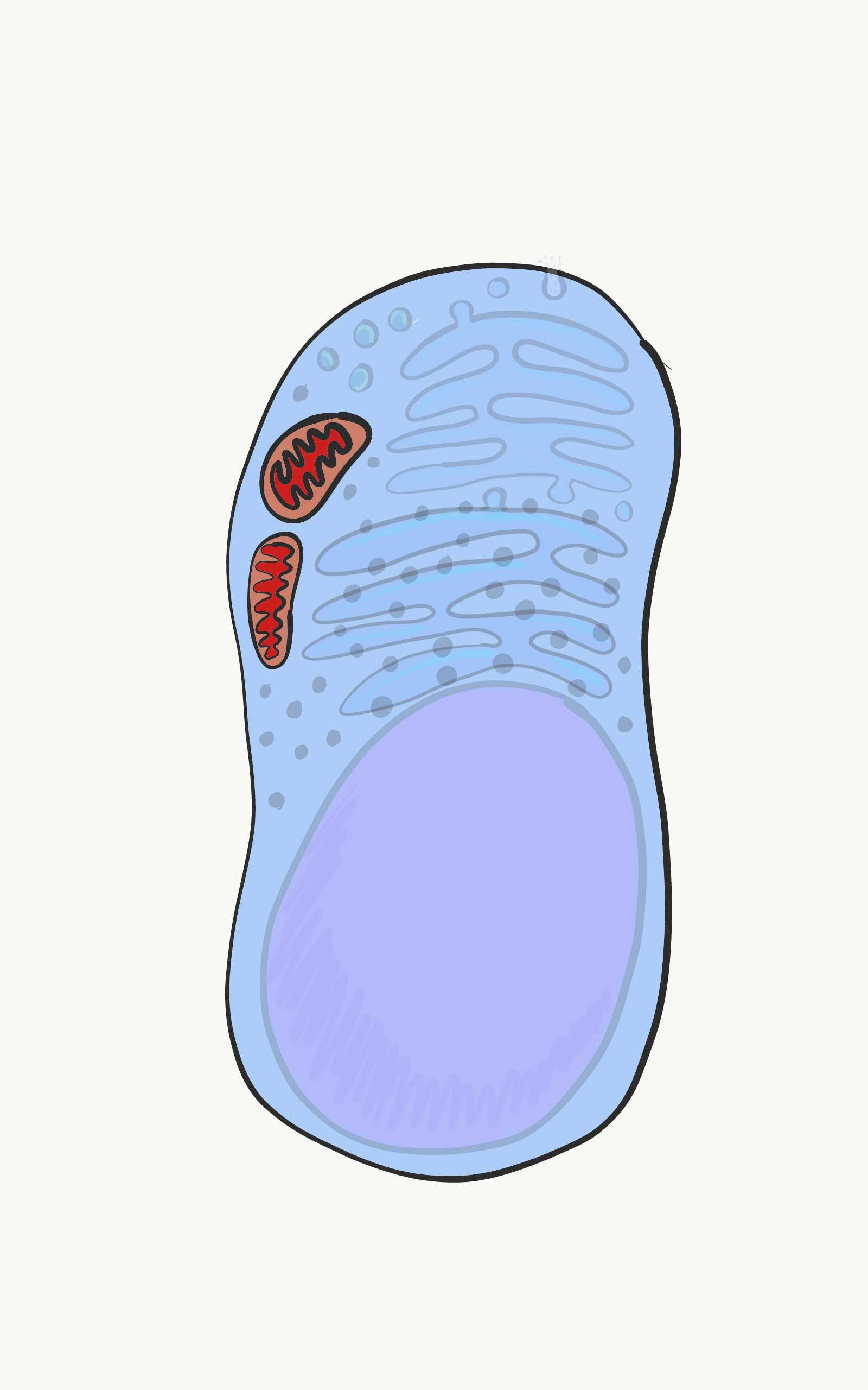
The mitochondria are where the majority of Adenosine Tri-Phosphate (ATP) is produced. ATP is made of Adenosine, plus three phosphate (PO43-) groups-- pay attention to the phosphate part, it is also a major component of bone, enamel, dentin and cementum. ATP powers almost all cellular processes, including the transcription and translation of mucous proteins within a salivary gland, the electrical signals sent by neurons in the tongue when food enters the oral cavity, and the contraction of myo-epithelial cells to cause salivation. Mitochondria burn glucose, using oxygen, and harness some of the energy released in the form of ATP.


Mitochondria are different from other organelles in that they contain a little bit of their own DNA, which is inherited just from mother. Mitochondria also contain two phopsholipid bilayer membranes, not 1 like other organelles. They use this extra membrane to generate ATP. Perhaps you covered glycolysis and the citric acid cycle before. The part to remember is that mitochondria use a proton (or H+) gradient, which makes the inside of mitochondria acidic, and therefore potentially toxic to the rest of the cell.

cell

Lysosomes

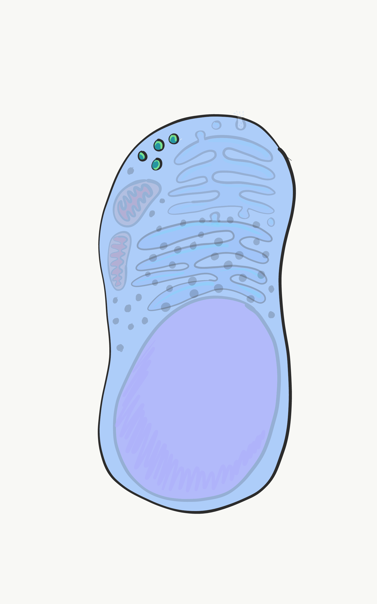
The lysosomes are small compartments surrounded by the same phospholipids as in the plasma membrane. Inside the lysosomes are acids and digestive enzymes that can be used to destroy stuff inside the cell when it wears out, or materials that the cell has gobbled up from outside (e.g. debris, bacteria).


When a cell dies and begins to break apart, neighboring cells are in danger of being damaged from the acids and enzymes wound within lysosomes. In the oral cavity, some epithelial cells will only hav a life-span of a few days before they wear out, so it will be very important for these cells to neutralize the acids and enzymes in their lysosomes first, in a process called apoptosis.

cell

Endoplasmic Reticulum

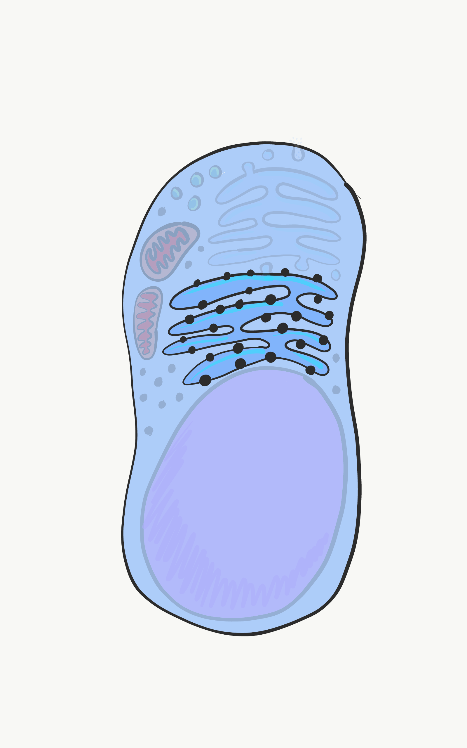
The ER is a series of interconnected tubes surrounded by a phospholipid bilayer-- similar to lysosomes, only bigger, more tubular, and not full of acid. The smooth Endoplasmic Reticulum (sER) is where cells produce lipids and store excess calcium. The rough Endoplasmic Reticulum (rER) is covered in ribosomes. Proteins made by these ribosomes wind up inside the rER, then travel to the golgi apparatus, and either wind up being secreted (such as mucous proteins) or stay within the plasma membrane (such as cell-junction proteins, or receptors for morphogen molecules).

cell

Golgi apparatus

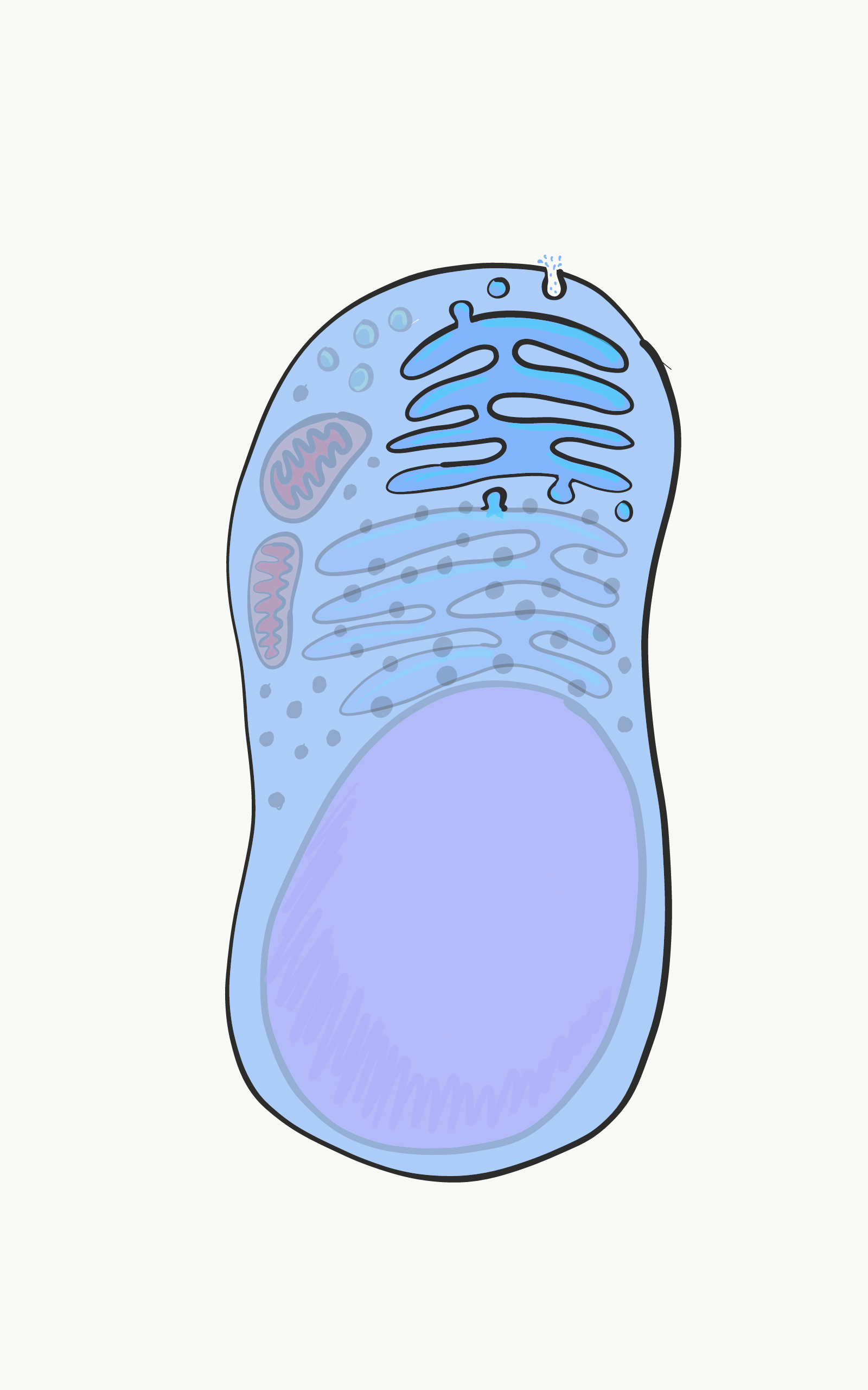
The Golgi apparatus is another set of tubes, similar to the rER. Vesicles shuttle proteins made in the rER to the Golgi apparatus, where the proteins are modified. Often, these proteins will have sugars attached to them, making them glycoproteins. New vesicles take these proteins to the plasma membrane, where they are either secreted or become a part of the plasma membrane. I will cover the role the secreted protein collagen plays in enamel and the periodontal ligament. I will also cover the shared role of the secreted glycoprotein fibronectin and the membrane-bound protein integrin have in healing damaged gingival tissue.

cell
By http://rsb.info.nih.gov/ij/images/, Public Domain CC0

Cytoskeleton

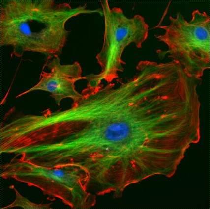
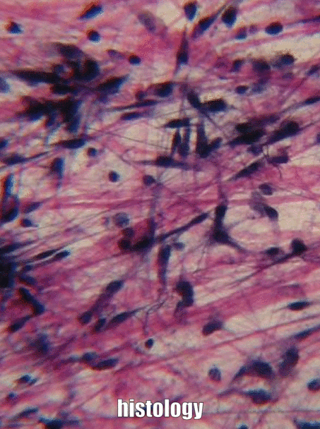
The cytoskeleton is a nextwork of long proteins within the cytoplasm. This network gives the cell its shape, the ability to change its shape, or to move. Shown here, these cells have their microtubules and actin filaments stained red and green. These proteins are not generally visible on more old-fashioned histology images.


Extra-Cellular Matrix

Ground Substance
Fibers
Extra-Cellular Matrix includes all the material found outside of cells. It is usually broken down into the two components to the left.

cell

Ground substance

Ground substance includes Extra-Cellular Fluid (ECF), which is the water and nutrients that was called plasma when it was inside of a blood vessel. Once fluid exits the blood and surrounds cells, it is called ECF. Unlike plasma, this fluid is held in place by proteins, glycoproteins, and polysacchrarides, forming a gel. This gel is ground substance. It doesn't look like much under a microscope, no more than if you looked really closely at some Jello. These proteins, glycoproteins and polysaccharides are made and secreted by cells (these cells probably have a lot of endoplasmic reticulum and golgi apparatus).


One of the glycoproteins found in ground substance is Fibronectin, which is a really long protein. Cell may recongize, bind to and move along fibronectin if they have the correct integrin protein on their plasma membrane. Therefore, fibronectin acts not only as a road along which cells travel, it is also a road map. Getting the right cells to the right place at the right time is very important both in healing and in development. In fact, what you learn about development will get re-used (recapitulated) in healing. If none of your patients have damage to their gingiva or teeth, you can skip this section. The rest, read on.

Another important molecule found in ground substance is a large polysaccharide called Hyaluronic Acid (HA). Like fibronectin, cells can bind to and travel over HA (using a different type of plasma membrane protein), which has applications in dentistry, such as helping cell of the gingiva stick to a dental implant and form a bacteria-resistant seal. We can't see fibronectin or HA without using some modern imaging tricks, which is why they are listed in as ground substance, and not in the next section, fibers.


Because cells can migrate over ground substance proteins, we say that ECM proteins function as a scaffold. Without scaffolds, tissues grow only from the outer edges. This is fine if speed is not important, such as when enamel and dentin are forming. However, in wound repair, it is optimal for a wound to heal everywhere at once, rather than from the edges. The body, therefore, often puts down some form of scaffolding first, such as a scab. In dentistry, artificial scaffolds can be created to help the body heal, based on our knowledge of the functions of ground substance.

In addition to their structural role as a scaffold, guiding cells to new locations, ground substance molecules can also provide cells with information. This information tells cells where they are located, and what they should be doing. For instance, when a stem cell binds to fibronectin, fibronectin can instruct the stem cell to express different genes and differentiate into a new type of cell, such as an odontoblast, and begin secreting dentin. Getting cells to the correct location is nice, but they need to know what to do when they get there. This happens during tooth formation, but also in reponse to tooth injury. As we learn more about how ground substance instructs stem cells, we get better at helping teeth repair themselves. I find it unfortunate that many textbooks gloss over ground substance as just the gelatinous material outside of a cell, which is why I have gone into it in more detail, and glossed over mitochondria.

Fibers

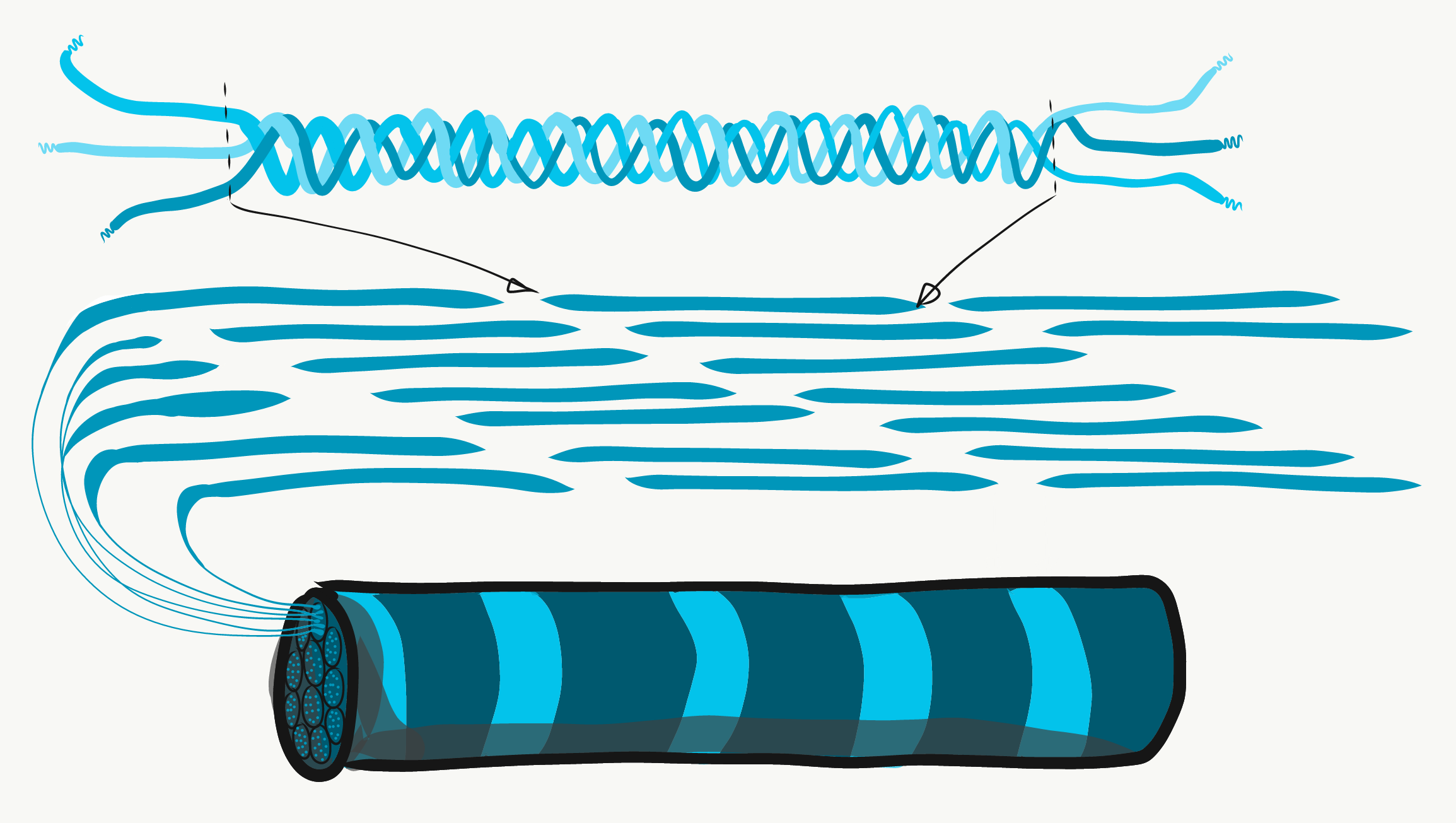
Three extracellular proteins were visible under a light microscope a century ago, so they were grouped together as fibers of the extra-cellular matrix. Like fibronectin an other ground-substance proteins, fibers are secreted by cells called fibroblasts.


collagen
Collagen is the strongest of the three. This fiber is made of 3 coiled α-helices, that are coiled and cross-linked, making a very strong macromolecule with the same basic shape as a rope. It is very strong if you pull on it from the ends, but not if you apply force from the side. Collagen fibers are found in regions of the oral cavity where the ability to resist force is important, such as in enamel or the periodontal ligament. In fact, its found throughout the human body, accounting for 25% of our proteins.


Reticlar fibers, which are not shown on the animated image above, look like a fine, spider web-like mesh under the microscope. Later it was discovered that reticular fibers are different shape of collagen, but they are still called by their own name, and receive equal footing with collagen on the list of ECM fibers. This web-like network of proteins isn't strong, but provides enough framework for blood cells to rest in organs like the spleen and lymph nodes.


Elastic fibers are thinner than collagen fibers, and often look like fine hairs in the ECM under a microscope. As their name suggests, these fibers can be stretched and then spring back to their original length. This is not something collagen can do. Elastic fibers are found in regions of the oral cavity that change shape during speech or swallowing, such as the soft palate.


mitosis
By Erin Rod - Own work, CC BY-SA 4.0

Mitosis

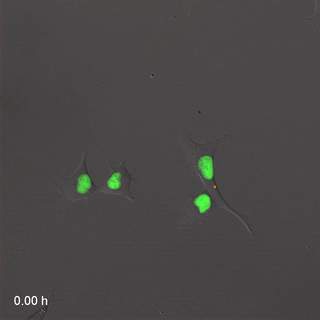
Cell division, or mitosis, the the process by which one cell makes a copy of itself, producing two identical daughter cells. Early in development, as we are growing from a single cell to a trillion cells, lots of mitosis occurs.

When a cell is not undergoing mitosis, it is said to be in interphase. This is the time where a cell might be doing its job, such as producing fibers for new ECM, or a cell might be preparing for mitosis. Before cell division can occur, a cell must have roughly double of everything. During mitosis, everything is divided in half between two new daughter cells. Not all cells are capable of mitosis-- in fact, most cells in an adult have differentiated and are performing tasks, they are too busy to reproduce. We say they have exited the cell cycle. To repair damage, tissues have stem cells which are capable of dividing. Cell division will produce two daughter cells, one daughter remains a stem cell, and the other differentiates into whatever cell is needed. A tissue will have a constant supply of stem cells, as long as the stem cells don't die before they can undergo mitosis. As we get older, our tissues don't heal as well because we have fewer stem cells.

Stem cells are named based on how many different types of cells they can potentially become. The uni-potent stem cells of the oral epithelium become keratinocytes, and only keratinocytes. The multi-potent Ecto-Mesenchymal Stem Cells turn into dentin, pulp, cementum and periodontal ligament. The omni-potent fertilized egg becomes every cell in a human, plus more.


cellcycle
by OpenStax is liscensed CC BY 4.0

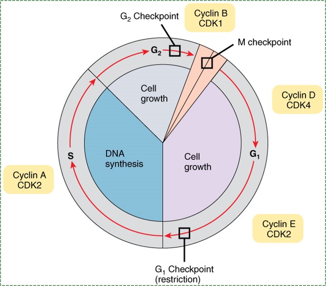
To go through interphase and prepare for another round of mitosis, cells go through a series of cell cycle checkpoints. This helps to regulate the timing of cell division, which in turn ensures that the correct amount of tissue growth occurs. The passage through cell cycle checkpoints is timed internally, by the cell itself. This involves slow, but regular phophosrylation of proteins called cyclins. Cyclins are transcription factors that can activate genes that allow progression through to a checkpoint. The speed of this process can be sped up or slowed down by external signals, such as growth factors. Growth factors are hormones that are secreted into the ground substance of a tissue. The density and stickiness of the ground substance influences how far the growth factor diffuses. If diffusion is limited, the growth factor only speeds up growth in a localized area. This is important in the formation of new organs, such as teeth. Other growth factors might spead over a wide area. This allows different organs to grow at roughly the same speed. Ensuring all the teeth are roughly the same size requires this pattern of growth factor release.


Mutation in a gene for a growth factor, or the receptor for a growth factor, can lead to a "gain-of-function": cells can receive a constant on-signal for passage through the cell cycle. We call these genes oncogenes. It takes just one bad copy of a receptor gene to gain a function. The cyclins and other genes that regulate the cell cycle, however, are called tumor supressor genes. For a cell to lose its ability to regulate cell cycle checkpoints, both copies of a tumor supressor gene must be mutated. In your car, it would take just one foot to step on the gas too hard, but you'd have to be missing both feet to not be able to hit the brakes. Usually, cancers only form when a single cell acquires mutations to both oncogenes and a pair of tumor supressor genes. Even then, there is another layer of protection that covered is the next section.

apoptosis
video by Birgit Janicke is in the public domain CC0

Apoptosis

All cells contain a group of cell-surface receptor proteins and intra-cellular enzymes that allow them to undergo programmed cell death, or apoptosis, when instructed. Programmed cell death is absolutely critical to multi-cellular life, which is an odd thing to say. Without it, as cells reached the end of their lifespan-- which for epithelial and blood cells is really short-- they would release the contents of their lysosomes and mitochondria. As you may recall, the contents of these organelles are highly acidic, which could damage or kill neighboring cells. If those neighboring cells died as a result, they too would release their lysosomal and mitochonrial contents, causing even more damage. When this happens in the human body it is called tissue necrosis. That's not all, a dead cell spews out DNA, and DNA is really long and stringy and tends to be very sticky. This can trap other cells and prevent them from migrating properly, which is great if you are trying to trap and kill bacteria, but otherwise not something you want to do to your neighboring cells.


Therefore, cells can be instructed to undergo apoptosis if they are reaching the end of their lifespan, if the immune system has determined them to be infected or cancerous, or just aren't needed anymore. Apoptosis ensures that before a cell dies, it neutralizes the pH of its lysosomes and mitochondria, and chops up its DNA into safe, small bits. During development, we will often see that more cells are produced than needed, and the extra cells are removed later in an organized fashion-- similar to the way construction of a large building involves building scaffolding first, and the scaffolding is removed towards the end of the project. The process of wound repair will also involve an over-production of cells followed by organized removal. Hopefully, this will seem logical by the time you finish this course, as we will see during wound repair, DNA instructions are turned on that haven't been used by cells since they were developing embryonically. If you want to sound fancy, and don't we all, you can say wound healing recapitulates (states again) embyronic development.


The process of apoptosis begins either with an internal or an external signal. For instance, when a cell's DNA becomes too mutated, or if there are an odd number of chromosomes during mitosis, this triggers apoptosis. Alternatively, a cell can be instructed to undergo apoptosis from an extracellular signal called a Tumor Necrosis Factor (TNF). A series of enzymes called caspaces are activated, which ultimately leads to the neutralization of acids, destruction of DNA, and cause the cell to explode into numerous small bits, which can be cleaned up by macrophages.

Cell junctions

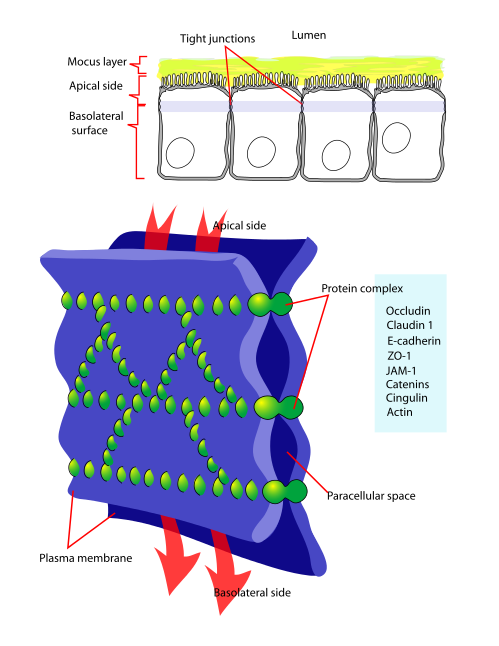
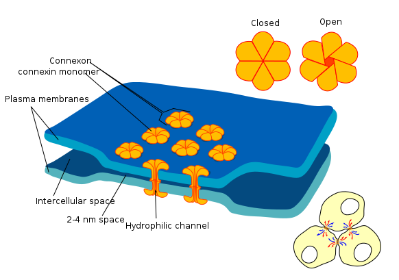
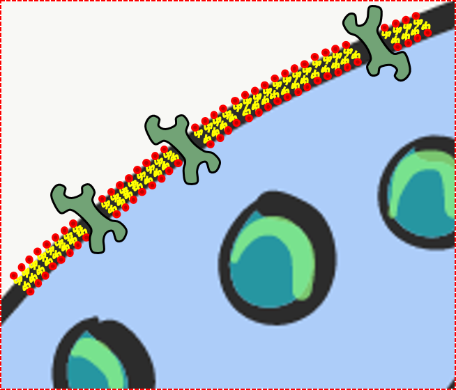
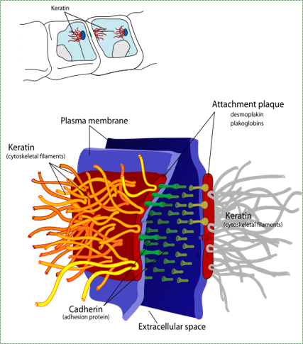
Cell-to-cell junctions
Cell-to-ECM junctions
Junctions are specialized groups of proteins on or near the cell surface that make connections to some other structure. These connections can be to other cells, or to the ECM.


junction
Own work By Mariana Ruiz LadyofHats - Public Domain CC0
Anchoring junctions (desmosomes) are strong connections between 2 cells. The anchoring junctions pair up and anchor the cytoskeleton of one cell to its neighbor. A large group of cells anchored together by these junctions will be much stronger as a group. A single twig breaks, but the bundle of twigs is strong --Tecumseh.


Hemi-desmosomes are half of a desmosome anchored to the ECM, such as the seal between the gingival epithelium and the non-cellular surface of a tooth. One of the many proteins in a desmosome is one called an Integrin. This protein can recognize and bind to proteins in the ECM such as fibronectin. When the Integrin protein of a cell connects to fibronectin, this not only anchors the cell's cytoskeleton to the ECM and anchor the cell in place, the integrin can also signal to the inside of the cell, allowing the nucleus to know what type of tissue the cell is located in.


Before a cell can migrate to a new location, it must first remove its anchoring junctions. During development, cells migrate to new locations and form new structures. During wound healing, stem cells will detach from their neighbors, migrate into the injured area, and begin cell division to create enough cells to fix the injury.


gap
Own work By Mariana Ruiz LadyofHats - Public Domain CC0
Tight junctions are smaller junctions between cells. Tight junctions completely encircle a cell, and create a water-tight seal between that cell and another cell. This serves to create barriers between one part of the body and another, allowing the cells to regulate what goes across and what does not. This also gives cells apical-to-basolateral polarity (or a difference between top and bottom), which is especially important to an epithelium. The apical side of an epithelial cell faces the lumen (hollow center) of an organ, while the basolateral side is closest to underlying connective tissue. Proteins synthesized on the rER can be sent to either the apical or basolateral portion of the plasma membrane. Once there, trans-membrane proteins of the apical side of the cell cannot diffuse to the plasma membrane on the basolateral side, because the ring of tight junctions blocks their movement.


gap
Own work By Mariana Ruiz LadyofHats - Public Domain CC0
Gap junctions, or connexons, are a group of proteins that form a gap between cells which can be opened or closed. This gap allows cells to communicate directly with one another. Beause of the way epithelial cells are connected to each other-- in a sheet-- this communication occurs across a plane. This is one way that cells know their position relative to one of the body's axes, and is a process called Planar Cell Polarity. PCP allows cells to know what direction they are facing in the body, ensuring that the structures they are forming are not only in the correct location, but in the correct orientation. For instance, this type of signaling allows teeth to form so that every tooth's lingual side is facing the lingual side, not facing the buccal, mesial or distal sides.